Introduction
Over recent decades, concern has been increasing regarding the declining human sperm count. Environmental, occupational, lifestyle, and dietary factors may affect reproductive health and contribute to decreases in semen quality [1,2]. Heavy metals adversely impact male reproductive health [3], even at relatively low levels of exposure [4], via either the disruption of the hypothalamic-pituitary axis or direct effects on spermatogenesis leading to reduced semen quality [5].
Lead is an environmental pollutant that can induce abnormalities in male reproductive function [6]. Significant negative correlations have been reported between semen quality and lead concentrations in the semen or blood [7]. Telisman et al. [4] found that blood lead levels in a group of Croatian men was positively associated with the percentage of pathological sperm. Lead levels in the sperm of Mexican men residing in urban areas were also demonstrated to show significant negative associations with sperm concentration, motility, viability, and normal morphology [8].
Oxidative stress, the major mechanism of lead toxicity, has been reported to occur as a result of reduced activity of antioxidant enzymes [9] and increased production of reactive oxygen species (ROS) [10], leading to lipid peroxidation and protein and nucleic acid oxidation [11]. Because the key constituents of the sperm cell membraneŌĆö polyunsaturated fatty acids and phospholipidsŌĆöare highly susceptible to oxidative damage, high concentrations of free radicals such as the superoxide anion, hydrogen peroxide, and nitric oxide directly damage the cells. This damage has been proposed as a possible etiology of idiopathic male infertility [12]. Accordingly, it has been suggested that antioxidants may protect body systems against various deleterious effects of lead [13].
In this regard, some believe that the adverse effects of toxic chemicals such as heavy metals may be mitigated by herbal remedies. Cichorium intybus L. is an herbaceous plant of the Asteraceae family that grows across much of Asia, Africa, and Europe. In the Ayurvedic and Unani systems of traditional medicine, C. intybus has been used to cure various ailments and has also been found to have wide-ranging applications in the food industry [14]. In Iranian folk medicine, C. intybus is known as kasni and has been used traditionally for its hepatoprotective, blood purifying, and male fertility-promoting properties [15-17]. Our previous study showed that C. intybus leaf extract improved reproductive parameters in male rats [18]. The whole extract of C. intybus has been reported to have anti-diabetic, antibacterial, immunostimulatory, and cardioprotective properties [19]. Phytochemical analyses have shown that C. intybus extract contains polyphenols such as chlorogenic acid, fructooligosaccharides, inulin, and caffeic acid derivatives [20]. Therefore, the present study was designed to evaluate the ability of C. intybus extract to protect against lead-induced oxidative stress and reproductive toxicity in adult male Wistar rats.
Methods
1. Preparation of C. intybus L. ethanolic extract
The leaves of C. intybus L. were collected from areas near Behbahan in Khouzestan, Iran, and authentication was conducted at the Botanical Systematic Laboratory, Department of Biology at Shahid Chamran University of Ahvaz in Iran. The dried C. intybus leaves (200 g) were ground to a fine powder and extracted using maceration with 96% ethanol (Merck, Darmstadt, Germany). The extract was filtered, and the solvent was evaporated using a rotary evaporator at 50┬░C. The extract was stored at 4┬░C until the time of use.
2. Experimental design
Fifty healthy 8-week-old male Wistar rats (body weight, 180ŌĆō200 g) were housed under standard conditions with regard to temperature (23┬░C ┬▒ 2┬░C) and light/dark cycle (12 hr/12 hr) and were given free access to standard food and water. The study protocol was approved by the Animal Ethics Committee of the Department of Biology at Shahid Chamran University of Ahvaz in Ahvaz, Iran. All experimental procedures were performed in accordance with National Institutes of Health guidelines for the care and use of laboratory animals (National Institutes of Health Publications No. 8023, revised 1978). The animals were randomly divided into five groups (n=10). The rats in the control group were gavaged with 1 mL of distilled water once daily. The rats of the second group were allowed to drink distilled water containing 0.1% lead acetate ad libitum and were given 1 mL of distilled water through gavage once daily. The rats in the remaining three groups were allowed to drink distilled water containing 0.1% lead acetate ad libitum and were treated, respectively, with 50, 100, and 200 mg/kg body weight of C. intybus extract via gavage once daily. The dose of lead acetate was chosen based on previously published studies and continued for 70 Consecutive days [21,22]. After 70 days, the animals were weighed and sacrificed under light ether anesthesia. Blood samples were collected in laboratory tubes via cardiac puncture and centrifuged at 3,000 rpm for 15 minutes, after which the reproductive organs were removed and weighed. The serum lead levels were estimated using the graphite furnace atomic absorption spectrometry method in the K Kimiay-e-Nab imiay-e-Nab analysis laboratory in Karaj, Iran.
3.Histological and histometric analysis
The right testes were excised, cleaned, and fixed in Bouin solution. Then, 5-╬╝m-thick paraffin sections were stained with hematoxylin and eosin for evaluation by light microscopy (Olympus, Tokyo, Japan). To measure the seminiferous tubule diameter, 2 perpendicular diameters of each cross-section were measured in 90 randomly chosen, nearly round cross-sections of the seminiferous tubules in each rat at a magnification of ├Ś 40.
4. Epididymal sperm analysis
The cauda epididymis was excised in a petri dish containing 1.0 mL of phosphate buffer (0.1 M, pH 7.4). The dishes were gently swirled for 10 minutes at 37┬░C to allow dispersion of the sperm cells in the solution. A 10-╬╝L aliquot of epididymal sperm suspension was placed on a hemocytometer and allowed to stand for 5 minutes; then, the sperm concentration was assessed and expressed in millions/mL of suspension. To gauge sperm motility, 10 ╬╝L of suspension was evaluated using a hemocytometer (Paul Marienfeld, Lauda-Konigshofen, Germany), and the percentages of motile and non-motile sperm were counted. To analyze sperm morphology, 40 ╬╝L of sperm suspension was mixed with 10 ╬╝L of 1% eosin Y for 45ŌĆō60 minutes at room temperature, and the percent of morphologically abnormal sperm was recorded.
5. Biochemical analysis
The left testes were homogenized in sodium phosphate buffer (0.1 M) at 4┬░C and centrifuged at 10,000 rpm for 15ŌĆō20 minutes at 4┬░C, and the supernatants were stored at ŌłÆ20┬░C until analysis. The activity levels of glutathione peroxidase (GPx) and superoxide dismutase (SOD) in the testicular homogenate were estimated using assay kits according to the manufacturerŌĆÖs instructions (Randox Laboratories, Crumlin, UK). The testicular levels of malondialdehyde (MDA) were determined using the thiobarbituric acid reaction and measured spectrophotometrically at 532 nm [23].
6.Hormonal assay
Serum levels of testosterone, luteinizing hormone (LH), and folliclestimulating hormone (FSH) were estimated using enzyme-linked immunosorbent assays (ELISA), using a commercial kit (AccuBind ELISA kit; Monobind Inc., Lake Forest, CA, USA) according to the manufacturerŌĆÖs instructions.
Results
1. Reproductive organ weight
Significant (p<0.05) decreases were observed in the weights of the testis, epididymis, seminal vesicle, and ventral prostate in lead acetate-treated rats compared with the control group (Table 1). Dose-dependent increases (p<0.05) were observed in the weights of the testis, epididymis, seminal vesicle, and ventral prostate in rats co-administered C. intybus extract relative to the lead acetate-treated rats (Table 1).
2.Histometric analysis
In the rats exposed to lead acetate, the seminiferous tubule diameter was significantly lower (p< 0.05) than in the control rats (Figure 1). The seminiferous tubule diameter was significantly higher (p< 0.05) in a dose-response manner in the lead acetate-treated rats co-administered C. intybus extract than in the rats administered lead acetate alone (Table 1).
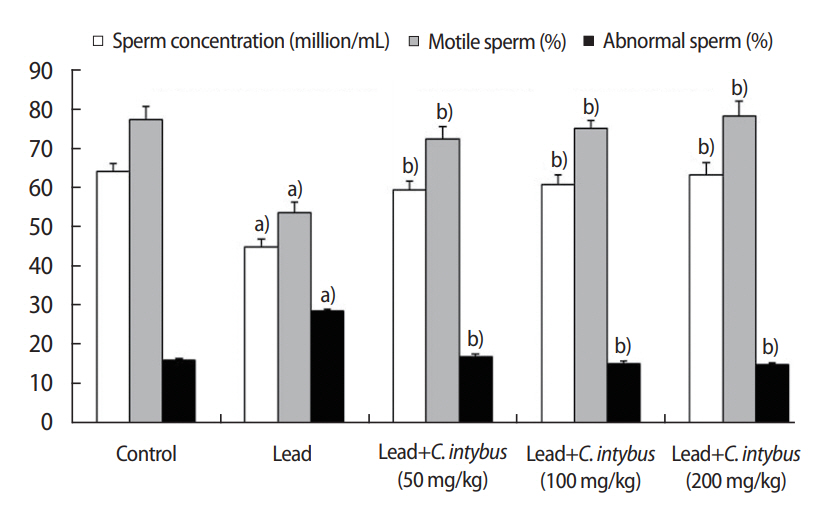
3. Epididymal sperm analysis
In the lead acetate-treated rats, relative to the control group, significant (p< 0.05) decreases were observed in the sperm concentration and the percent of motile sperm. A higher percentage of sperm with abnormal morphology (p< 0.05) was also observed in the lead acetate-treated rats (Figure 2). The coadministration of C. intybus extract significantly improved (p<0.05) sperm parameters in a dose-response manner compared with the lead acetate-treated rats (Figure 3).
4. Biochemical analysis
In the rats treated with lead acetate, the testicular levels of MDA were significantly higher (p< 0.05) than in the control group. Moreover, the activity levels of SOD and GPx were significantly lower (p< 0.05) in the testes of the rats exposed to lead acetate (Table 2). In a dose-dependent manner, MDA levels were shown to decrease and the levels of activity of SOD and GPx were shown to increase significantly (p< 0.05) in the rats coadministered C. intybus extract (Table 2).
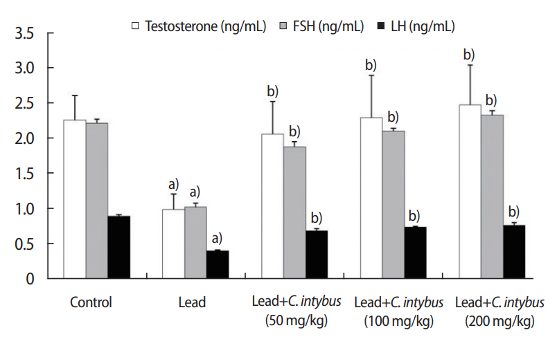
5.Hormonal assay
Compared with the control group, significant (p< 0.05) declines were seen in serum testosterone, FSH, and LH levels in the lead acetate-treated rats (Figure 4). In the rats co-administered C. intybus extract, significantly (p< 0.05) higher levels of testosterone, FSH, and LH were observed compared with the lead acetate-treated group, and these results were dose-dependent (Figure 4).
Discussion
Traditional herbal remedies are a complementary therapy used to enhance fertility and to improve semen parameters in infertile men. The aim of the present study was to evaluate the potential protective effects of C. intybus leaf extract on lead-induced oxidative reproductive toxicity in male Wistar rats. C. intybus is known in different areas of the world for its numerous properties in traditional medicine and is used in Iranian folk medicine for its supposed fertility-enhancing properties. Given that exposure to heavy metals can trigger lipid peroxidation and cause oxidative stress damage, antioxidants may be an effective defense mechanism against lead-induced toxicity. In this study, the effects of co-administration of C. intybus leaf extract on reproductive and oxidative status parameters were evaluated in male rats exposed to low levels of lead acetate. The results showed that coadministration of C. intybus mitigates testicular toxicity and improves male reproductive function in lead-treated rats.
Our findings demonstrated that exposure to lead acetate adversely affects reproductive parameters in adult male rats. The toxic effects of lead on male reproductive function may be direct (via affecting spermatogenesis and sperm function) or indirect (by disturbing the hypothalamic-pituitary-testicular axis) [22,24]. Weighing reproductive organs is widely considered to be a valuable screening tool [25]. In the present study, the weight of the testis was significantly decreased in lead-treated rats, a finding that aligned with earlier reports [26-28]. Crucially, the disruption of spermatogenesis has been shown to reduce the mass of differentiated spermatogenic cells and lead to a decrease in the testis weight [29]. In addition, the weights of the testes and male accessory reproductive glands are known to be positively correlated with testosterone levels, meaning that the adequate bioavailability of testosterone is essential for the structural and functional integrity of reproductive organs [30]. Therefore, a reduction in serum testosterone levels due to a decrease in androgen biosynthesis led to significant decreases in the weights of the testis, prostate, and seminal vesicle in the lead-treated rats.
However, the co-administration of C. intybus leaf extract resulted in dose-related increases in epididymal sperm count in the lead-treated rats. Epididymal sperm count is considered to be one of the most sensitive tests used to evaluate testicular function, sperm production, and male fertility [31]. The decreases in serum testosterone levels explain the reduction in sperm count even in light of the finding of epidydimal weight loss in lead-treated rats [32]. Thus, lead toxicity could cause a reduction in sperm count and an increase in the percentage of abnormal spermatozoa, resulting in reduced fertility [33]. The increases in sperm count and the percent of normal morphological sperm in lead-treated rats therefore indicate that treatment with C. intybus improves and enhances the fertilizing capacity of semen. In addition, increased levels of MDA and decreased activity of SOD and GPx in the testes of adult rats show that exposure to lead results in testicular oxidative stress. Our findings are consistent with the results of previous reports [26,34]. The excessive generation of ROS overwhelms the bodyŌĆÖs antioxidant defense and leads to oxidative stress, which has been reported to be a major mechanism underlying lead toxicity [35]. The binding of lead to sulfhydryl groups or metal cofactors of antioxidant enzymes such as SOD and GPx reduces the activity of these enzymes [36]. Higher levels of ROS have been found to be associated with impaired sperm motility, decreased concentration, and altered morphology [37]. The generation of free radicals results in lipid peroxidation in the plasma membrane, which is known to play a significant role in the etiology of sperm dysfunction [38].
This study showed that C. intybus leaf extract improves testicular oxidative status by decreasing the levels of lipid peroxidation and increasing the activity of SOD and GPx in the testes. In vitro studies have indicated that the constituents of C. intybus, especially from the red varieties of the plant, may possess anti-free radical activity [39]. Previous studies have shown that C. intybus extract could protect against oxidative stress and hepatotoxicity induced by paracetamol or carbon tetrachloride [40-42]. Phytochemical analyses have shown that the phenolic compounds that are present in large amounts in C. intybus alcoholic extract are important contributors to its antioxidant properties [14,43]. The protective properties of phenolic compounds against cellular oxidative damage stem from their hydrogen donating ability [44]. Blokhina et al. [45] showed that phenolic compounds hinder the diffusion of free radicals, restrict the peroxidative reaction, and reduce membrane fluidity and thereby stabilize cell membranes [45]. However, this is not only true of phenolic compounds; rather, the sugars of chicory preparations, especially sucrose and fructans, have also been proposed to act as radical scavengers in plant cells [46]. Consequently, the present study showed that the co-administration of C. intybus extract attenuated lead-induced testicular oxidative stress and toxicity by reducing lipid peroxidation and increasing the activity of antioxidant enzymes. This co-administration thereby improved reproductive efficiency in lead-treated adult male rats. However, whether C. intybus extract can reverse the reproductive effects of lead-induced toxicity requires further study. Our findings support the beneficial effects of C. intybus on male reproductive health and its traditional use for the treatment of male infertility. However, the study was limited by the lack of available information regarding the safety of C. intybus extract. Further investigations are needed to evaluate the toxicity of C. intybus extract and the therapeutic efficacy of its bioactive constituents.