Effect of electromagnetic field exposure on the reproductive system
Article information
Abstract
The safety of human exposure to an ever-increasing number and diversity of electromagnetic field (EMF) sources both at work and at home has become a public health issue. To date, many in vivo and in vitro studies have revealed that EMF exposure can alter cellular homeostasis, endocrine function, reproductive function, and fetal development in animal systems. Reproductive parameters reported to be altered by EMF exposure include male germ cell death, the estrous cycle, reproductive endocrine hormones, reproductive organ weights, sperm motility, early embryonic development, and pregnancy success. At the cellular level, an increase in free radicals and [Ca2+]i may mediate the effect of EMFs and lead to cell growth inhibition, protein misfolding, and DNA breaks. The effect of EMF exposure on reproductive function differs according to frequency and wave, strength (energy), and duration of exposure. In the present review, the effects of EMFs on reproductive function are summarized according to the types of EMF, wave type, strength, and duration of exposure at cellular and organism levels.
Introduction
Humans in modern society are exposed to an ever-increasing number of electromagnetic fields (EMFs) generated from the production and supply of electricity, television (TV) sets, personal computer (PC), radio communication, and mobile communication. Since the 1960s, when the biological hazard of EMF exposure was studied in the Soviet Union, the safety of humans exposed to EMFs both at home and during occupational activities has become an important issue in public health. The biological effect of EMFs is currently under debate and still a controversial issue. In the present review, the effects of EMFs on reproductive function are summarized according to the types of EMFs and duration of exposure at the cellular and organism levels.
Types of EMF and frequency of exposure
Humans in modern society are exposed to various kinds of EMFs. Extremely low frequency (ELF)-EMFs have 3 to 30 Hz frequencies and are generated from military communication. The EMFs to which humans are most frequently exposed are the 50 to 60 Hz super low frequency (SLF) EMFs generated from power cables for industrial and household electrical supplies and electronic goods. Very low frequency (VLF) EMFs with 3 to 30 kHz frequency are generated from PC monitors or TV sets. EMFs from TVs or PCs have a 6.25 µT intensity with a 20 kHz frequency [1]. The radio frequency (RF) EMFs generated from mobile phones, cordless phones, and broadcasting towers have frequencies of hundreds of MHz. All these EMFs are non-ionizing radiation, which do not have energy to release electrons from orbit. EMFs have a wave character in short frequency and act as a magnetic field in long frequency. The strength of the electric field and magnetic field is measured in units of kV/m and µT, respectively. Household electronic goods can produce a 4 µT EMF and EMF ranges from 0.01 to 1 µT inside and outside of house [2]. The strength of SLF-EMFs is dependent on the electrical current and distance from the conductor. Therefore, SLF-EMFs are the highest near the power cable and decrease rapidly by distance.
Cellular mechanism for EMF induced toxicity
At the cellular level, an increase in free radicals and [Ca2+]i may mediate the effect of EMFs and lead to cell growth inhibition, protein misfolding, and DNA breaks. EMFs can disrupt Ca2+-dependent cell signaling. How does EL-EMF exposure affect signal transduction in cells? In human leukemia T-cell line Jurkat cells, a 50 Hz, 0.5 mT EMF was found to increase Ca2+ levels, blocking the effect of cholera toxin and the protein tyrosine kinase inhibitor genistein [3]. In thymic lymphocytes, Ca2+ influx increased during mitogen-activated signal transduction when exposed to a 60 Hz, 22 mT EMF [4], suggesting the modulating role of EMF on regulation of Ca2+ channels. RF EMFs of 900 and 872 MHz may enhance chemically induced reactive oxygen species (ROS) production, resulting in secondary DNA damage in human SH-SY5Y neuroblastoma cells [5,6]. Also in vivo experiments revealed the increased oxidative stress caused by a 900 MHz EMF, leading to endometrial histopathologic impairment in rats [7]. In prostate cancer cells, ROS induced by a 60 Hz sinusoidal EMF inhibited cell growth by apoptosis and arrested the cell cycle [8]. An RF EMF of 2,450 MHz exposure caused rearrangement of DNA segments and breakage of DNA in testes [9]. In another report, 1,800 MHz of EMF induced DNA breaks in human fibroblasts and rat granulosa cells in comet assay [10]. Similarly, an RF-EMF of 1,800 MHz induced DNA damage in Chinese hamster lung cells [11]. In addition, an RF EMF of 900 MHz and 1.7 GHz induced DNA breakage in cauda epididymal spermatozoa and embryonic stem cells in mice [12,13]. Some investigators have reported changes in protein folding by EMF. Changes in the structural fluctuation of tuna myoglobin protein was induced by EMF at the mobile phone frequency of 1.95 MHz, indicating RF EMFs as a potential risk for protein misfolding [14]. Heat-shock proteins (HSPs) were also increased by EMF exposure. In human endothelial cell line EA.hy926, HSP27 was activated by 900 MHz GSM non-thermal exposure [15]. HSP70 is induced by exposure to SLF (<300 Hz) EMFs [16]. Interestingly, HSP70 has non-thermal (EMF domain) and thermal (temperature domain) stress response promoter binding sites [17,18], suggesting that HSP70 is highly sensitive for EMF. In hepatocellular carcinoma, cell proliferation was inhibited with mitotic spindle disruption by a 27.12 MHz of RF EMF [19]. EMFs have been suggested as a cancer treatment tool with gamma irradiation. When a human breast cancer xenograft was treated with EMF and gamma irradiation at the same time, inhibitory effects on growth, angiogenesis, and metastasis were higher than in xenografts treated with gamma irradiation alone [20] (Figure 1).

Summary of the effects of electromagnetic fields at the cellular level. EMF, electromagnetic field; N, nucleus; ER, endoplasmic reticulum; M, mitochondria.
Human diseases related to EMF exposure
Alterations in biomarkers following EMF exposure have been reported through in vitro and in vivo experiments using animal cells and animals, respectively. Most of what is known on the correlation between human health and EMF exposure has been drawn from epidemiological studies. The results on the hazards of EMF exposure are contradictory, leaving the conclusion unclear to date [21-23]. Currently, the biological hazard of EMF exposure is understood to be different according to frequency, type of wave, and strength (energy) of the EMF. Importantly, some people are very sensitive to certain types of EMFs. The effect of EMF exposure could be different at various toxicological endpoint levels according to the route and duration of EMF exposure and target human subjects. Therefore, the possible body or cellular functions susceptible for EMF exposure should be suggested according to the type of frequency, wave, and strength of EMF, and the safety guidelines for EMF exposure should be made according to this criteria.
Possible human diseases related with EMF exposure obtained from epidemiological studies include the life threatening diseases such as leukemia in children and adults [24,25], brain cancer in adults [26], Lou Gehrig's disease [27], depression [28], suicide [29], and Alzheimer's' disease [30]. Recently, EMFs were reported to cause DNA damage and neurological diseases at much lower levels than those proscribed by international safety guidelines. Most recently, the Bioinitiative Report (http://www.bioinitiative.org/) has noted that the current safety guidelines for EMF exposure are not sufficient and should be revised based on data from various toxicological tests [31].
Changes in the reproductive endocrine system by EMF exposure
There are many studies on the casual relationship between SLF-EMF exposure and pineal gland function [32]. Although still under debate, EMF exposure can affect the secreting activity of the pineal gland in several animal species as does the light. Indeed, static exposure to SLF-EMFs can affect cyclic secretions of melatonin in several species [33]. In Long-Evans rats exposed to a circularly polarized 50 Hz EMF for 6 weeks, the pineal and circulating melatonin levels decreased [34]. In cows, a 60 Hz EMF exposure for 4 weeks (16 hours per day) altered circulating melatonin and prolactin levels and the estrous cycle [35,36]. In an adult Djungarian hamster, a 60 Hz EMF exposure acutely affected the pineal and circulating melatonin levels [37]. SLF-EMF exposure directly affects the pineal gland, deteriorating the biological effect of melatonin [38]. Melatonin regulates the pulse of LHRH in the hypothalamus, influencing gonadotropin FSH and LH. Eventually, this can alter the production of gonadal sex steroids, resulting in changes in the reproductive cycle [39,40]. In cows, exposure to 60 Hz SLF-EMF at 30 µT for 24-27 days did not alter the progesterone levels but shortened the estrous stage [41]. RF-EMF exposure can affect ACTH, GH, TSH, FSH, and LH in the pituitary [42]. Most EMF-induced hormonal changes are mediated by the thermal effect of EMFs. In contrast, long-term exposure to RF-EMFs did not have a cumulative effect on the endocrine, serological, or immunological parameters [43].
Reproductive toxicity of EMF in females
Reproductive toxicity of EMF exposure has been studied in both sexes at various endpoints [44]. Undoubtedly, reproduction is under control of the nervous system and endocrine system. In female mice, neuroendocrinological alteration was believed to be a prime cause of loss of fertility with aging [45,46]. In female mice exposed to a 20 kHz saw tooth EMF generated from a TV set or PC for 6 weeks after weaning, the estrous cycle was extended [47]. In cows, similarly, a 60 Hz, 30 µT EMF 16 hours per day extended the estrous cycle [35]. Because extension of the estrous cycle can decrease total ovulation opportunities in females during their fertile period of life, decrease in fecundity can be expected. In mouse follicle cultures, exposure to a 33 Hz SLF-EMF at 5-day intervals resulted in defects in follicle growth. In contrast, the follicles exposed to a 50 Hz EMF continued growth. 33 Hz or 50 Hz SLF-EMF exposure for 3 days inhibited the antrum formation of follicles cultured in vitro [48]. Together, the estrous cycle regulated by ovarian steroids might be much more sensitive to EMF exposure than the fetal development and feto-maternal interaction. In contrast, in female rats a 10 kHz, 0.2 mT sine wave EMF does not affect the estrous cycle [1], suggesting that the effect of EMFs on the estrous cycle differs according to the frequency, energy, and animal species. In adult Wistar female rats exposed continuously to a 50 Hz SLF-EMF for 3 months, the weights of the uterus and ovaries, progesterone levels, and estrogen levels in relation to the varying periods of the estrous cycle were not significantly altered [49].
When ovariectomized female Sprague-Dawley rats were exposed to a 1,439 MHz time division multiple access (TDMA) EMF for 4 hours per day for 3 days, there were no differences in uterine wet mass or serum estradiol level, suggesting no estrogenic activity related to high frequency EMFs used in cellular phones [50]. When female rats were exposed to a 900 MHz EMF for 30 min/day for 30 days, endometrial apoptosis and oxidative stress were increased [51]. Also, as an in vivo experiment, increased oxidative stress by a 900 MHz EMF led to the endometrial histopathologic impairment in rats [7].
Reproductive toxicity of EMFs in males
A code division multiple access (CDMA) EMF of 848.5 MHz has no significant effect on the body, testicular, or epididymal weights, or the sperm count or apoptosis of germ cells in adult Sprague-Dawley rats [52]. Similarly, in adult male Sprague-Dawley rats exposed 30 minutes per day, 5 days a week for 4 weeks to a 900 MHz EMF, the testes weight, the testicular biopsy score count, and the percentage of interstitial tissue out of the entire testicular tissue were not significantly changed. However, the diameter of the seminiferous tubules, the mean height of the germinal epithelium, and serum total testosterone levels were significantly decreased in the EMF group. Plasma LH and FSH levels had not significantly changed following EMF exposure [53]. In prepubertal male rats (age of 5 weeks) exposed to a 1.95-GHz wide-band CDMA signal for 5 hours per day, 7 days a week for 5 weeks, neither the body weight gain or weights of the testis, epididymis, seminal vesicles and prostate, nor the number of sperm in the testis and epididymis had changed. Of note, the testicular sperm count was significantly increased without abnormalities of sperm motility or morphology, suggesting a positive effect of W-CDMA on spermatogenesis [54]. However, at a higher frequency, specifically 2.45 GHz, an EMF induced a decrease in the Leydig cell number and increase of apoptosis-positive cells in the seminiferous tubules of Wistar rats [55]. Exposure to an RF EMF of 2,450 MHz resulted in rearrangement of DNA segments and breakage of DNA in the testis [9]. An RF EMF of 900 MHz and 1.7 GHz induced DNA breakage in mouse cauda epididymal spermatozoa and embryonic stem cells [12,13]. Taken together, the RF-EMF generated by a mobile phone might be harmful to male reproductive function.
In male mice, a 60 Hz EMF did not decrease the body or testes weights but significantly increased germ cell death and abnormality in the seminiferous tubules. A 60 Hz EMF at 0.5 mT increased the TUNEL-positive spermatogonia, indicating the potentiation of DNA fragmentation, but in flow cytometry, the cell survival was not significantly altered [56]. Similarly, 60 Hz EMFs of 14 and 200 µT also induced the apoptosis of spermatogenic cells in mice [57]. In human sperm, the acrosome reaction was not altered by a 900 MHz RF-EMF from mobile phone at 2.0 W/kg strength for 1 hour though the sperm morphology was changed together with a decrease in sperm-hemizona binding, indicating that the RF-EMF exposure can decrease sperm fertility [58]. In contrast, acute exposure to a sinusoidal SLF-EMF (50 Hz, 1 mT) did not affect boar sperm viability and morphology during the first hour of incubation. SLF-EMF treated spermatozoa showed resting intracellular Ca2+ levels significantly lower than those recorded for controls. As a consequence, MF-SLF exposed sperm displayed a reduced motility, a modest reactivity when co-incubated with solubilized zonae pellucidae, and a reduction in oocyte penetrating ability. After 2 or 4 hours of incubation, signs of morphological damage appeared on the plasma membrane and at the acrosome, and decreased the fertilization rate. A 1 mT EMF decreased sperm function in vivo [59,60]. In rabbits, a 50 Hz SLF-EMF was found to be able to alter sperm motility and decrease their viability [61]. Together, SLF-EMFs negatively influence spermatozoa first by impairing cell Ca2+ homeostasis then by affecting sperm morphology and function. In contrast, the positive effect of SLF-EMFs on sperm was also reported. In human sperm, a 50 Hz, 5 mT square wave SLF-EMF increased sperm motility within 3 hours, and this effect was sustained for 21 hours. In contrast, a 50 Hz, 5 mT sine wave EMF and 50 Hz, 2.5 mT square wave SLF-EMF did not affect sperm motility. Therefore, the positive effect of a SLF-EMF on sperm motility was dependent on the type and strength of the EMF [62].
The toxicity of EMF exposure to the next generation has also been reported. Male rat offspring exposed to 60 Hz, 1 mT SLF-EMF from gestation day 13 through postnatal day 21 showed a decrease in the number, diameter, area, and volume of seminiferous tubules, the height of seminiferous epithelium, and the number of Leydig cells, but connective tissues, vasculature, plasma testosterone levels, Sertoli cells, the length of seminiferous tubules, and gonadosomatic index remained unchanged. This suggests that gestational and lactational exposure to SLF-EMFs has adverse effects on the testis development [63]. In contrast, when pregnant rats were exposed to a 60 Hz, 500 µT SLF-EMF for 21 hours per day from gestation day 6 to postnatal day 21, spermatogenesis and the fertility of male offspring were not significantly different from the control [64].
Developmental toxicity of EMFs
The developmental toxicity of EMF exposure has been studied at various endpoints [44]. The effect of EMF exposure on implantation and fetal development was reported. When female mice were superovulated and mated following exposure to a 50-Hz EMF at 0.5 mT 4 hours per day, 6 days a week for 2 weeks, the number of blastocysts was significantly decreased together with an increase in DNA fragmentation. This suggests that EMF exposure in the preimplantation stage could have detrimental effects on embryo development [65]. In swine, cleavage of fertilized eggs in the oviduct was delayed under 50 Hz, 0.75 mT EMF exposure 4 hours before ovulation, suggesting that SLF-EMFs negatively affect early embryo development [60]. When pregnant mice were exposed to a 50 Hz, 20 mT sine wave EMF during gestation day 0 to 17, embryonic survival, sex ratio, and embryonic malformation were not significantly changed, but the height and body weight of offspring were significantly increased [66]. When males and females were exposed to a 50 Hz, sine wave EMF at 5.0 mT for 9 weeks and 2 weeks before copulation, respectively, the fertility of both gametes and fetal development were not affected [67]. In mice, when the dams were exposed to a 20 kHz saw tooth EMF at 6.5 µT for 8 hours per day during pregnancy, fetal abnormality was not increased [68]. The potential hazard of EMF exposure to the dam and fetus was reviewed in [1]. There was a positive relationship between occupational monitor labor during pregnancy and the natural abortion rate [69-71]. Epidemiological studies on birth defects and abortions in pregnant women working in offices revealed that the EMF generated from a computer monitor can negatively affect human reproduction [69,72]. However, none of these kinds of reports have been verified by experiments [73]. Importantly, the reproductive hazard of EMF exposure in non-pregnant young women has not been studied well. Together, the effect of SLF-EMF exposure on embryonic development is still controversial, but some negative effects of EMFs have been reported in some animal models. Thus, it would be better to avoid or minimize casual exposure to EMFs during pregnancy.
Conclusions and perspectives
Through in vitro and in vivo studies, EMF exposure was found to alter the reproductive endocrine hormones, gonadal function, embryonic development, pregnancy, and fetal development (Table 1, Figure 2). These effects were different according to the frequency, duration of exposure, and strength of EMFs. Humans in modern society cannot avoid various kinds of EMFs during household and occupational activities, but should be aware of the biological hazard of EMFs. The effort to avoid EMF exposure and techniques to protect or relieve EMF radiation are required to preserve our reproductive potential.
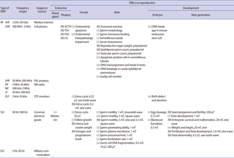
Effects of EMF on mammalian endocrine system and reproduction
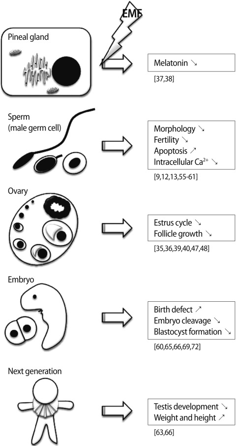
Summary of the effects of electromagnetic fields (EMFs) on reproduction. ↗, increase; ↘, decrease or inhibition.
Notes
No potential conflict of interest relevant to this article was reported.