Comparative analysis of conventional in vitro fertilization and intracytoplasmic sperm injection in patients with polycystic ovarian syndrome, tubal factor infertility, and unexplained infertility whose partners exhibit normal semen parameters: a retrospective study of sibling oocytes
Article information

Abstract
Objective
This study compared the outcomes of conventional in vitro fertilization (IVF) and intracytoplasmic sperm injection (ICSI) in patients with polycystic ovarian syndrome (PCOS), tubal factor (TF) infertility, and unexplained infertility whose partners had normal semen parameters.
Methods
This retrospective study included 360 couples diagnosed with infertility involving PCOS (n=157), unexplained infertility (n=140), and TF infertility (n=63). Sibling oocytes were randomly assigned to undergo ICSI or conventional IVF insemination. The fertilization rate and embryo morphology were evaluated as outcomes.
Results
Retrieved cumulus-oocyte complexes from patients with PCOS (2,974), unexplained infertility (1,843), and TF infertility (844) were split and inseminated by conventional IVF and ICSI respectively. In comparison to the ICSI method, the conventional IVF approach was linked to a significantly higher fertilization rate in groups with PCOS (68.81% vs. 77.49%), unexplained infertility (67.62% vs. 78.84%), and TF issues (69.23% vs. 78.63%) (p<0.05). The proportion of embryos with grade A produced by the conventional IVF method was significantly higher than that produced using the ICSI method in the PCOS and unexplained infertility groups (p<0.05). Additionally, the percentage of grade B embryos produced with the ICSI method was significantly higher than that produced with the conventional IVF method in PCOS patients (p=0.002).
Conclusion
Our results indicated that the conventional IVF method was associated with higher zygote production and a higher proportion of grade A embryos when all infertile groups were evaluated together. Thus, ICSI is not suggested for patients with these causes of infertility if their partner has normal semen parameters.
Introduction
Infertility is a global issue, and the World Health Organization (WHO) anticipates that it will become the third most serious disease after cancer and cardiovascular disease during this century [1]. The WHO estimates that approximately 8% to 10% of couples worldwide are affected by infertility problems. Polycystic ovarian syndrome (PCOS), tubal factors, and unexplained infertility are among the primary causes of infertility [2]. PCOS is a common endocrine disorder with an incidence rate of about 4% to 8%, though it can be as high as 25% in some populations [3]. It has been noted that over 70% of women with PCOS exhibit normal-gonadotrophic anovulation [4]. These women often present with polycystic ovaries, hirsutism, oligomenorrhea or amenorrhea, and anovulatory cycles. PCOS is also linked to metabolic abnormalities, insulin resistance, and an elevated risk of cardiovascular disease and type 2 diabetes [5]. Due to oligo-ovulation or anovulation, patients with PCOS often require assisted reproductive technology (ART) to achieve pregnancy [6].
Unexplained infertility is a commonly reported diagnosis in infertility centers. Unfortunately, due to the lack of sufficient diagnostic tests to identify definitive factors of infertility, the cause remains unknown in some couples, a condition referred to as unexplained infertility [7]. It is estimated that 15% to 30% of infertile couples will be diagnosed with unexplained infertility [8]. The National Institute for Health and Care Excellence (NICE) guidelines on infertility suggest that women with unexplained infertility should attempt to conceive through natural intercourse for a period of 2 years. If pregnancy has not been achieved after this period, conventional in vitro fertilization (IVF) and/or intracytoplasmic sperm injection (ICSI) are recommended as the next effective treatment options [9].
The fallopian tubes are crucial for capturing the ovulated egg and facilitating the transport of sperm and the embryo. When these tubes malfunction, it can lead to tubal factor infertility, which is a leading cause of female infertility [10]. Most commonly, tubal factor infertility is caused by occlusion and peritoneal pathology, which result in adhesions. Approximately 30% to 35% of infertile women are affected by this condition. There are two prevalent treatments for tubal factor infertility: tubal surgery and IVF. IVF offers several advantages, including higher success rates per cycle, being less surgically invasive, and allowing couples to attempt conception immediately after a diagnosis of tubal factor infertility is made. However, there are drawbacks to IVF, such as the risk of ovarian hyperstimulation, the high cost, and the increased likelihood of multiple gestations [11].
As ART has evolved over the past several decades, it has become a treatment option for nearly all forms of infertility. In terms of patient conditions, the majority undergo either conventional IVF or ICSI. Although the NICE recommends ICSI primarily for cases of male factor infertility or following unsuccessful IVF attempts [9], the use of ICSI instead of conventional IVF has been growing, even among couples without male factor infertility, increasing from 15% in 1996 to 67% in 2012 [12]. While one previous review did not report differences in pregnancy rates between IVF/ICSI in couples with female factor infertility [13], another review suggested that ICSI improves the fertilization rate and decreases the likelihood of complete fertilization failure in couples with unexplained infertility [14]. At our clinic, we perform conventional IVF and also utilize ICSI for infertile patients with an adequate number of oocytes, aiming to boost fertilization rates and reduce the need to repeat ART cycles due to failure. Although there is no definitive evidence that ICSI is superior to conventional IVF in terms of reproductive outcomes in certain cases of infertility, the purpose of this study is to compare the outcomes of conventional IVF and ICSI (specifically fertilization rates and embryo quality) in patients with PCOS, tubal factor infertility, and couples with unexplained infertility where the male partner has normal semen parameters.
Methods
1. Patients
Data were collected from 360 couples who visited the Afzalipour Infertility Research and Treatment Center in Kerman for IVF/ICSI treatment between May 2016 and April 2021. These patients were experiencing infertility issues, including PCOS, unexplained infertility, or tubal factor infertility. Three cohorts consisting of 157, 140, and 63 patients with similar clinical parameters were formed corresponding to the PCOS, unexplained infertility, and tubal factor groups, respectively. All patients underwent conventional IVF and ICSI procedures. The male partners all had normal basic semen parameters according to the WHO standards [15]. The Ethics Committee and Institutional Review Board of Kerman University School of Medical Sciences approved this retrospective randomized study (IR.KMU.AH.REC.1401.013), and informed consent was obtained from all participants included in the study.
Women (n=157) aged 18 to 40 years with a diagnosis of PCOS were randomly selected based on the 2003 Rotterdam criteria. The diagnosis was made according to at least two of the following three criteria: (1) ovarian dysfunction (oligo/anovulation); (2) excess androgens, including clinical or biochemical hyperandrogenism; (3) exclusion of other causes of androgen excess (e.g., congenital adrenal hyperplasia and androgen-secreting tumors) or ovulatory disorders (e.g., Cushing syndrome) [16]. Total serum testosterone levels were measured to evaluate hyperandrogenism. Polycystic ovaries were identified by ultrasonography, defined as an ovary with a volume greater than 10 cm3 or an ovary containing more than 12 follicles measuring 2 to 9 mm in diameter. Exclusion criteria for this study included severe systemic diseases (such as cardiovascular, liver, or kidney diseases), benign or malignant gynecological tumors (including cervical cancer, endometrial tumor, and ovarian tumor), and allergy to gonadotropins.
A cohort of 140 couples, aged between 20 and 40 years and experiencing unexplained infertility, was evaluated. All women participating in the study exhibited normal ovulatory cycles, uterine cavities, and fallopian tube patency, as confirmed by hysterosalpingography. Cycles were excluded from the study if, 12 days following the commencement of follicle-stimulating hormone (FSH) administration, either (1) fewer than three follicles measuring 17 to 18 mm in diameter were present, or (2) more than 20 follicles were observed, including the leading three follicles, accompanied by a serum estradiol level exceeding 1,600 Ci/mmol, to prevent the risk of ovarian hyperstimulation syndrome.
Tubal factor infertility was diagnosed in women (n=63) aged 18 to 40 years through laparoscopy or laparotomy due to hydrosalpinx, medial or lateral occlusion. These women had normal ovarian function, regular menstrual cycles, luteal phase progesterone levels exceeding 15 nmol/L, and normal concentrations of thyroid-stimulating hormone, prolactin, and free thyroxine. Women with PCOS were excluded from the study.
2. Ovarian stimulation protocol
All women with PCOS included in the study received a daily dose of highly purified human menopausal gonadotropin (HP-hMG) for 3 consecutive days. Ovarian stimulation was performed using either recombinant FSH or HP-hMG following a gonadotropin-releasing hormone (GnRH) antagonist protocol. The clinician determined the initial dosage of HP-hMG and the GnRH analog. When ultrasound scans revealed three leading follicles measuring 17 to 18 mm in diameter, an injection of either human chorionic gonadotropin (HCG; 5,000 to 10,000 IU) or triptorelin (0.2 mg) was administered. Cumulus-oocyte complexes (COCs) were retrieved 36 hours after the HCG injection, using a 17-gauge single-lumen needle while the patient was under general anesthesia. The COCs were immediately harvested from the follicular fluid. For each patient, the COCs were divided into two groups: one for IVF and the other for ICSI. Both conventional IVF and ICSI procedures were performed for all patients included in the study. The number of retrieved oocytes, as well as the number of metaphase II (MII), metaphase I (MI), germinal vesicle (GV), and degenerated oocytes, were compared between the two groups.
3. Fertilization procedure
COCs (n=5,661) were collected from three categories of infertile couples: those with PCOS, unexplained infertility, and tubal factor infertility. For ICSI procedures, COCs were first enzymatically denuded of their surrounding cumulus layers using hyaluronidase, followed by mechanical denudation through pipetting under a stereomicroscope. The denuded oocytes were then assessed for integrity and maturity using an inverted microscope (Nikon TE 300). Mature oocytes (MII) were identified by the extrusion of the first polar body. In the ICSI groups, MII oocytes underwent microinjection as described by Van Landuyt et al. [17]. Conventional IVF involved the insemination of COCs with progressively motile sperm at a concentration of 0.1×10⁶/mL. Both conventional IVF and ICSI were performed for each couple included in this study. Fertilization was confirmed 16 to 18 hours post-insemination by the presence of two pronuclei within the zygotes. As outcomes, the fertilization rate and embryo morphology, were evaluated.
4. Embryo culture and quality assessment
Zygotes were placed in a 25 μL droplet of Sage one-step culture medium (CooperSurgical Fertility Companies) and cultured until day 3 at 37 °C in an atmosphere containing 5% CO2. Embryo quality was assessed according to the criteria established by Hill et al. [18], which consider the size of blastomeres and the percentage of fragmentation. Embryos were classified into four groups: grade A (blastomeres of equal size with no fragments); grade B (blastomeres of unequal size with less than 10% cytoplasmic fragments); grade C (blastomeres of unequal size with more than 50% fragmentation); grade D (blastomeres of unequal size with severe fragmentation and large black granules). The patterns of embryo fragmentation were compared between these groups.
5. Statistical analysis
The normality of the data distribution was analyzed using the Kolmogorov-Smirnov test. Variables that were parametric and normally distributed were analyzed using the paired Student’s t-test for comparisons between the IVF and ICSI groups, and one-way analysis of variance was conducted for age, fertilization rate, and embryo quality. To compare means among the different infertile groups, the Tukey honest significant difference post hoc test was employed. Nonparametric data, including the duration of infertility, number of retrieved oocytes, and the counts of MII, MI, GV, and degenerated oocytes, were compared using the Kruskal-Wallis test, followed by the Dunn multiple comparison test. A p-value <0.05 was considered to indicate statistical significance. Data were presented statistically as mean/number or percentages, with the standard deviation indicated. Statistical analyses were performed using SPSS ver. 20 software (IBM Corp.).
Results
Patients with PCOS had a lower mean age (28.55±3.89) compared to those with unexplained infertility and tubal factor infertility (31.87±4.06 and 31.97±4.14, respectively; p=0.01). The duration of infertility in the tubal factor infertility group was significantly shorter (3.73±3.27 years) than in the PCOS group (5.79±3.08 years) and the unexplained infertility group (5.21±3.36 years; p=0.001) (Table 1).
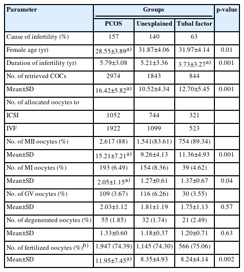
Comparison of characteristics of the three types of infertility
The total number of retrieved COCs in the PCOS, unexplained, and tubal factor groups was 2,974, 1,843, and 844, respectively. The PCOS group exhibited a significantly higher mean number of retrieved oocytes than the unexplained and tubal factor groups (16.42±5.82, 10.52±4.34, and 12.70±5.45, respectively; p<0.001). In all three groups, COCs were divided between ICSI and conventional IVF procedures. Within the PCOS group, 1,052 COCs underwent ICSI, while 1,922 COCs were used for conventional IVF. In the unexplained infertility group, 744 COCs were allocated to ICSI and 1,099 to IVF. For the tubal factor group, 321 COCs were assigned to ICSI and 523 to conventional IVF.
The percentages of mature (MII), immature (MI), and fertilized oocytes in the PCOS group were significantly higher than those in the other infertile groups (p=0.001, p<0.05, and p=0.002, respectively). There were no statistically significant differences in the rates of GV and degenerated oocytes among the groups (p>0.05) (Table 1). Table 2 presents a comparison of the fertilization rates and embryo quality outcomes between ICSI and conventional IVF methods across the three groups studied.

Comparison of fertilization rate and embryo quality between the ICSI and conventional IVF groups in patients with PCOS, unexplained infertility, and tubal factor infertility
In the PCOS group, 642 zygotes were produced via ICSI, while 1,305 zygotes were generated through conventional IVF. The fertilization rate for conventional IVF was significantly higher than that of ICSI (77.49%±21.57% vs. 68.81%±23.88%, p=0.008). Furthermore, a significantly higher proportion of grade A embryos were produced in the conventional IVF group compared to the ICSI group (16.40% vs. 9.81%, p=0.001). The ICSI group, however, yielded a greater percentage of grade B embryos (54.83%) than the conventional IVF group (46.05%, p=0.002). No statistically significant differences were found in the percentages of grade C and D embryos between the conventional IVF and ICSI methods (p>0.05).
In patients with unexplained infertility, the fertilization rate in the conventional IVF method (78.84%±22.85%) was significantly higher than ICSI (67.62%±23.51%) (p=0.03). Moreover, a higher percentage of grade A embryos was produced in the conventional IVF group (10.10%) than in the ICSI group (7.81%) (p=0.04). However, the percentages of grades B, C, and D embryos were similar between the two methods (p>0.05).
Similarly, in women with tubal factor infertility, the fertilization rate was higher with the conventional IVF method (78.63%±19.49%) than with the ICSI method (69.23%±22.15%) (p=0.01). Additionally, the proportion of grade D embryos was greater in the conventional IVF group (19.02%) than in the ICSI group (10.60%) (p=0.02). There were no statistically significant differences in the percentages of grades A, B, and C embryos between the two groups (p>0.05).
As indicated in Table 2, the fertilization rate and embryo quality resulting from both IVF and ICSI did not differ significantly among patients with PCOS, unexplained infertility, and tubal factor infertility (p>0.05). However, the proportion of grade A embryos produced by IVF was higher in the PCOS group (16.40%) than in the unexplained infertility and tubal factor groups (10.10% and 10.33%, respectively) (p=0.0001).
Discussion
We studied three groups of infertile women—those with PCOS, those with unexplained infertility, and those with tubal factor infertility—who underwent IVF and ICSI as treatments for infertility. The primary objective of any IVF/ICSI program is to harvest a substantial number of mature oocytes while avoiding the risk of ovarian hyperstimulation syndrome. This risk is particularly pronounced in patients with PCOS, as these women tend to be more sensitive to exogenous stimulation than women without PCOS [19]. In our study, the number of retrieved, MI, and MII oocytes was higher in the PCOS group than in the unexplained and tubal factor infertility groups. These findings align with a previous study that reported a greater number of retrieved oocytes in the PCOS group than in the tubal factor group. However, that study also noted a lower fertilization rate in the PCOS group relative to the tubal factor group. Additionally, it suggested that the outcomes of IVF/ICSI might be similar between these two groups [20], which contradicts the results of our study. The higher number of fertilized oocytes and grade A embryos observed in the PCOS group in our research could be attributed to the higher number of oocytes retrieved from women with PCOS compared to those from the other two groups.
The data showed that in patients with PCOS and those with unexplained infertility, the fertilization rate and the proportion of grade A embryos were higher in the IVF group compared to the ICSI group. Additionally, a larger number of grade B embryos were obtained from the ICSI group in patients with PCOS. In a retrospective study involving women aged 40 years or older with unexplained infertility, the fertilization rate was found to be higher with IVF than with ICSI [21]. This finding appears to be at odds with those of a previous study—a systematic review and meta-analysis on unexplained infertility—which suggested that ICSI was superior to IVF in terms of increasing the fertilization rate per retrieved oocyte. This review included 11 studies of sibling oocytes from women with unexplained infertility, which were randomly allocated to either ICSI or IVF. The patients selected for this meta-analysis had an average number of retrieved oocytes ranging from 10.8 to 16.3, a range similar to that of our study [14].
The duration of infertility in women with tubal factor infertility was shorter than in those with PCOS and unexplained infertility. tubal factor infertility tends to be diagnosed more quickly than unexplained infertility, and the initiation of ART in these women occurs earlier than in those with PCOS and unexplained infertility, leading to a shorter duration of infertility in women with tubal factor infertility. In this group, fertilization and embryo fragmentation rates (grade D) were higher in IVF compared to ICSI. This finding aligns with a previous study by Aboulghar et al., which analyzed women with tubal factor infertility and divided them into two groups. Their results indicated that the fertilization rate per retrieved oocyte was higher in the IVF group than in the ICSI group. They noted that in the ICSI method, only MII oocytes are used, whereas in IVF, MI oocytes may mature in the culture media and subsequently become fertilized, thus potentially improving the fertilization rate per retrieved oocyte [22]. Moreover, despite the oocyte sources, culture medium, and laboratory conditions being identical in both methods, the differences between the two groups may also be attributed to the invasive nature of the ICSI method.
Our results indicated that the IVF method was associated with higher zygote production and a greater proportion of embryos with grades A and D when all infertile groups were evaluated together. A potential reason for these differences could be the mechanical injury to oocytes and sperm caused by the invasive procedure of microinjection in the ICSI technique. Conversely, these results highlight the importance of natural sperm selection. These findings align with those of some researchers who have indicated that fertilization rates in the IVF group are comparable to, or even higher than, those in the ICSI group [23-25]. It appears that the insemination technique used in our study improved embryo quality, as evidenced by the higher number of grade A embryos in the IVF method across all non-male factor infertility cases. Thus, our study supports the notion that these three factors of infertility do not confer a putative advantage over ICSI when partners have normospermic semen. In contrast to our findings, Lee et al. [26] reported no difference in embryo quality between ICSI and IVF in a group of couples undergoing oocyte split insemination who had mild male factor infertility, tubal factor infertility, or unexplained infertility.
The deleterious effects of microinjecting oocytes on embryo quality remain inconclusive. Various reports have indicated that the quality of embryos resulting from ICSI can be comparable to, lower than, or higher than those derived from IVF [27-29]. Frattarelli et al. [30] observed that the morphology of IVF embryos was superior to those from ICSI, regardless of semen parameters. Their findings indicated increased embryo fragmentation and a reduced number of non-fragmented grade A embryos with the ICSI method [30]. Conversely, another study reported similar fertilization rates between IVF and ICSI in patients without male factor infertility, with a higher incidence of grade A embryos in the ICSI group [27]. Our results suggest that the micromanipulation involved in the ICSI process is associated with increased embryo fragmentation. There are few studies that investigate the mechanical damage caused during denudation and micromanipulation for microinjection. Despite advancements in ICSI technology, mechanical micromanipulation still carries a 5% to 19% risk of oocyte degeneration [31,32].
The fertilization rate and embryo quality resulting from ICSI were comparable among patients with PCOS, unexplained infertility, and tubal factor infertility. These findings indicate that ICSI should not be prioritized for these three types of infertility. Additionally, our results imply that ICSI does not contribute to an increase in fertilization rate and embryo quality in cases of PCOS, unexplained infertility, and tubal factor infertility when male factor infertility is not present. This conclusion aligns with the recommendations of the Practice Committee of the American Society for Reproductive Medicine, which states that there is no supportive evidence for the use of ICSI in non-male factor infertility [33]. Furthermore, due to its invasive nature, ICSI is an expensive and time-consuming technique that requires additional equipment and skilled technicians.
In the absence of evidence-based guidelines, some clinics routinely use ICSI for women, regardless of the cause of infertility, based on the belief that ICSI may reduce the likelihood of fertilization failure [34]. However, the findings of this study indicate that ICSI does not result in higher fertilization rates or improved embryo quality compared to IVF in the treatment of women with PCOS, tubal factor infertility, or unexplained infertility when their partners have normal semen parameters. Therefore, we do not recommend the use of ICSI for patients with these types of infertility when their partner's semen analysis is normal. Additionally, women undergoing ICSI should be informed about the potential impact of this procedure on embryo quality. Further research is necessary to determine whether ICSI offers any benefits over IVF or if its use may adversely affect embryos from patients with non-male factor infertility.
Notes
Conflict of interest
No potential conflict of interest relevant to this article was reported.
Author contributions
Conceptualization: RH, SA (Sanaz Alaee). Formal analysis: SA (Sareh Ashourzadeh), RH, RK, SS (Saeed Shokri). Investigation: SA (Sareh Ashourzadeh), SS (Somayyeh Safari). Supervision: SA (Sanaz Alaee). Writing-original draft: SA (Sareh Ashourzadeh). Writing-review & editing: SS (Somayyeh Safari), SA (Sanaz Alaee). Approval of final manuscript: SA (Sareh Ashourzadeh), SS (Somayyeh Safari), RH, RK, SS (Saeed Shokri), SA (Sanaz Alaee).