Clin Exp Reprod Med Search

CLOSE


Clin Exp Reprod Med > Volume 47(3); 2020 > Article
Golestanpour, Javadi, and Sheikhha: Association of GRIA1 polymorphisms with ovarian response to human menopausal gonadotropin in Iranian women

Abstract

Objective

Glutamate ionotropic receptor AMPA type subunit 1 (GRIA1) is a subunit of a ligand-gated ion channel that regulates the secretion of follicle-stimulating hormone (FSH) and luteinizing hormone (LH) by controlling the release of gonadotropin-releasing hormone. Few studies have investigated the association between the GRIA1 gene and human infertility. This study evaluated the association of the GRIA1 rs548294 C > T and rs2195450 G > A polymorphisms with the ovarian response to human menopausal gonadotropin (HMG) in Iranian women.

Methods

One hundred women with histories of at least 1 year of infertility were included. On the second day of menstruation, patients were injected with HMG; on the third day, blood samples were collected. After hormonal analysis, the GRIA1 rs548294 C > T and rs2195450 G > A genotypes of samples were identified via the restriction fragment length polymorphism method, and on day 9, the number of follicles was assessed via ultrasound.

Results

For the GRIA1 rs548294 C > T and rs2195450 G > A single nucleotide polymorphisms, the subjects with CT and GG genotypes, respectively, displayed the highest mean FSH level, LH level, and number of follicles on day 9 of the menstrual cycle (p < 0.05). Significant positive correlations were observed between LH and FSH (p < 0.01), LH and follicle count (p < 0.01), FSH and age (p < 0.05), follicle count and age (p = 0.048), and FSH and follicle count (p < 0.01).

Conclusion

This study showed a significant relationship between GRIA1 polymorphisms and ovarian response to the induction of ovulation. Therefore, determining patients’ GRIA1 genotype may be useful for improving treatment and prescribing suitable doses of ovulation-stimulating drugs.

Introduction

Infertility refers to the failure of couples to conceive after at least 1 year of unprotected coitus, and it affects 13%–18% of couples [1,2]. Female factors are responsible for nearly 30% of all infertility cases. The main reason for female infertility is ovulation dysfunction [3], which impacts around 40% of women suffering from infertility [4]. Hormone secretion is crucial to the initiation of ovulation; therefore, impairment of hormone pathways can lead to ovulation dysfunction [5,6].
An appealing treatment option for women with dysfunctional ovulation is ovulation induction, which also increases the quality and quality of oocytes for use with assisted reproductive techniques [7-9]. Several ovulation induction drugs exist, the most common of which are human menopausal gonadotropin (HMG) and clomiphene citrate [10,11]. Ovarian hyperstimulation and multiple births are the most important side effects of these medications [12-14]. The use of newer methods and a more comprehensive understanding of patients’ genetic background can play an important role in reducing the side effects of these drugs.
Follicle-stimulating hormone (FSH) [15] and luteinizing hormone (LH) [16] are the hormones most crucially involved in the regulation and initiation of ovulation [13,17]. In women, the reduced secretion of LH or FSH interferes with the reproductive cycle. Medications that regulate the levels of FSH and LH are widely used to facilitate assisted reproductive techniques [9,18]. The present study is of particular importance because if we can recognize the gene variations that affect ovulation (especially the induction of ovulation), we can use this information to design advanced infertility treatment.
The glutamate ionotropic receptor AMPA type subunit 1 (GRIA1) gene, which encodes a subunit of a ligand-gated ion channel, is located on chromosome 5q33.2 and spans 20 exons [19,20]. In humans, a strong association between the GRIA1 rs548294 C > T and rs2195450 G > A polymorphisms and migraine risk has been demonstrated in many case-control studies [19,21,22]. Moreover, GRIA1 regulates FSH and LH secretion by controlling the release of gonadotropin-releasing hormone [23]. Very few studies have investigated the association between the GRIA1 gene and infertility in humans, and most related studies have been performed in cattle [23]. In addition, studies in cattle have shown that the GRIA1 polymorphism results in the replacement of serine with asparagine. This reduces the release of gonadotropin-releasing hormone, resulting in defective ovulation [24]. Considering the findings regarding the relationship between GRIA1 genetic variants and ovulation, the purpose of the present study was to evaluate the association of the GRIA1 rs548294 C > T and rs2195450 G > A polymorphisms with the ovarian response to ovarian stimulation treatments in Iranian women.

Methods

1. Study population

This cross-sectional study was performed on 100 infertile women (age: range, 20–35 years) who had been referred to the Abortion Research Center at Yazd Reproductive Sciences Institute. The Institutional Ethics Committee of Yazd Reproductive Sciences Institute approved the study, and written informed consent was obtained from all participants. All patients were treated with long-protocol ovulation induction, and on the second day of menstruation, patients were injected with HMG. On the third day of the menstrual cycle, a blood sample was collected in a simple tube with no additive for serum extraction and evaluation of LH and FSH levels. A blood sample was also collected in an ethylenediamine tetraacetic acid (EDTA)-containing tube for the extraction of DNA and the determination of the genotype with regard to GRIA1 polymorphisms. On the ninth day of the menstrual cycle, the number of follicles was evaluated via ultrasound. Patients with a history of pelvic surgery or ovarian cyst were excluded. All patients were candidates for in vitro fertilization (IVF) for various reasons (male factor infertility, uterine malformations, or unexplained infertility) and had experienced at least 1 year of infertility.

2. Genotyping

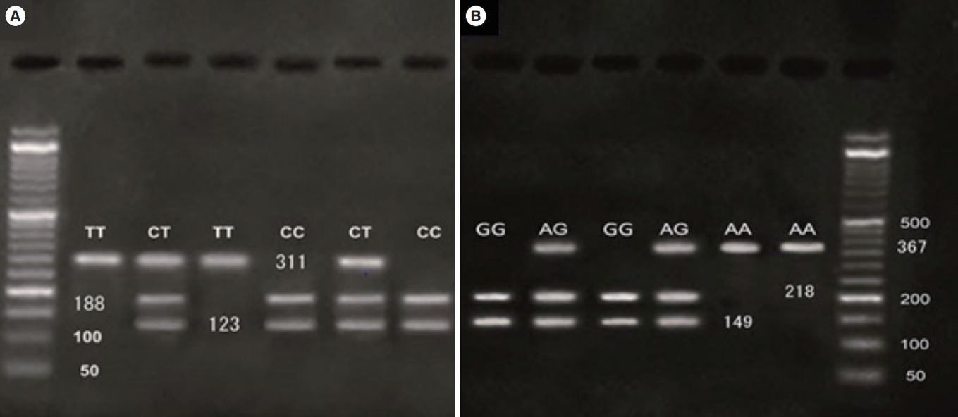
Genomic DNA was obtained from whole blood using the Qiagen QIAamp DNA Blood Mini Kit (Cat No. 51105; Qiagen, Hilden, Germany). The quality and quantity of extracted DNA were quantified via an agarose gel and spectrophotometry (optical density 260/280), respectively. The GRIA1 rs548294 G>A and rs2195450 C>T genotypes were assessed via the polymerase chain reaction (PCR)-restriction fragment length polymorphism technique. PCR of the GRIA1 rs548294 C>T and rs2195450 G>A polymorphisms was conducted using the following primers: for the GRIA1 rs548294 C>T polymorphism, a forward primer of 5´-AGATGAAGAAACAGAGGTC-3´ and a reverse primer of 5´-CCCCAGGTACTATTCAAAG-3´; and for the rs2195450 G >A polymorphism, a forward primer of 5´- TCTAAGAGGAGGGGGCAAGG-3´ and a reverse primer of 5´- GCTTGGTAGATGGTGCTTGA-3´.
The GRIA1 rs548294 C > T and rs2195450 G > A polymorphisms were identified by digesting the PCR products with the restriction endonucleases MwoI and TaqI, respectively. An agarose 3% gel and electrophoresis were applied, and the results were immediately visualized under ultraviolet illumination for assessment of the genotypes of the specimens (Figure 1).

3. Statistical analysis

Statistical analysis was performed using SPSS ver. 18.0 (SPSS Inc., Chicago, IL, USA). Differences in allele and genotype frequencies among the patients were analyzed via the chi-square test and analysis of variance. Logistic regression was used to calculate odds ratios and 95% confidence intervals. Two-tailed p-values of less than 0.05 were considered to indicate statistical significance.

Results

1.Hormonal analysis

The mean LH and FSH levels by GRIA1 rs548294 C>T and rs2195450 G > A polymorphism genotype are presented in Tables 1 and 2, respectively. For the GRIA1 rs548294 C > T single-nucleotide polymorphism (SNP), the frequencies of genotypes CT, CC, and TT were 48%, 29%, and 23%, respectively. For the GRIA1 rs2195450 G > A SNP, the frequencies of genotypes AG, GG, and AA were 20%, 75%, and 5%, respectively. The results indicate a significant relationship between the mean levels of LH and FSH and the GRIA1 rs548294 C > T SNP; specifically, the mean LH and FSH levels associated with the CT genotype were higher than those associated with the other two genotypes (p = 0.000 for both LH and FSH). These outcomes also show a significant relationship between the mean levels of LH and FSH and the GRIA1 rs2195450 G > A SNP, with the GG genotype associated with higher levels of both LH and FSH than the other two genotypes (p = 0.029 and p = 0.000, respectively).

2.Number of follicles at day 9 of the menstrual cycle

The mean number of follicles at day 9 of the menstrual cycle by GRIA1 rs548294 C > T and rs2195450 G > A polymorphism genotype are shown in Table 3. Of the 91 patients for whom the mean number of follicles was assessed, for the GRIA1 rs548294 C > T SNP, the number of cases with genotypes CT, CC, and TT were 44, 27, and 20, respectively; for the GRIA1 rs2195450 G > A SNP, the number of cases with genotypes AG, GG, and AA were computed as 18, 68, and 5, respectively. A significant relationship was observed between the mean number of follicles at day 9 and both GRIA1 rs548294 C > T and rs2195450 G > A SNPs. Specifically, regarding the GRIA1 rs548294 C > T SNP, the mean number of follicles was higher in subjects with the CT genotype than in those with the other two genotypes (p = 0.002). Regarding the GRIA1 rs2195450 G > A SNP, the mean number of follicles was higher in subjects with the GG genotype than in those with the other 2 genotypes (p = 0.000).

3. Correlation between LH, FSH, and number of follicles

The Pearson correlation analysis showed significant positive correlations between LH and FSH levels (r = 0.504, p < 0.01), LH levels and number of follicles (r = 0.611, p < 0.01), FSH levels and age (r = 0.207, p < 0.05), number of follicles and age (r = 0.192, p = 0.048), and FSH levels and number of follicles (r = 0.438, p < 0.01). However, no significant correlation was found between LH levels and age (r = 0.162, p = 0.107) (Table 4). These results illustrate the interactions among FSH and LH levels, age, and number of follicles.

Discussion

Ovarian dysfunction occurs in approximately 15%–25% of infertility cases, and the response to treatment is typically very satisfactory when ovarian dysfunction is the only cause of infertility [25]. Briefly, successful treatment in these individuals depends on careful examination and identification of the underlying cause of ovulatory dysfunction. A reliable method to predict ovarian response to different ovarian stimulation methods would be extremely helpful in determining the prognosis of these methods with regard to pregnancy. Thus, it is necessary to consider the genetic backgrounds of individuals that may lead to varied responses to different doses of ovarian stimulation drugs.
To the best of our knowledge, this experiment was the first that assessed the association between GRIA1 rs548294 C > T and rs2195450 G > A polymorphisms and ovarian response to ovarian stimulation treatments in Iranian women. The hormonal analysis showed that, for the GRIA1 rs548294 C > T and rs2195450 G > A SNPs, respectively, the CT and GG genotypes were associated with higher mean FSH and LH levels than the other genotypes. These polymorphisms in GRIA1 lead to the replacement of serine with asparagine. GRIA1Asn has a weaker affinity for glutamate than GRIA1Ser. This replacement decreases the secretion of gonadotropin-releasing hormone, leading to defective ovulation. In cows, GRIA1 polymorphisms result in the reduced release of gonadotropin-releasing hormone and a decreased response to hormone treatment. Due to the crucial role of GRIA1 in regulating FSH and LH secretion, it is important to consider the genotypes associated with increased levels of these hormones.
The number and size of follicles are important factors in the success of assisted reproductive methods [26]. Many factors, including genetic variants, age, and hormones, contribute to follicle quality [27]. In this study, comparison of the mean number of follicles at day 9 of the menstrual cycle showed that, similarly to the mean LH and FSH levels, the number of follicles in subjects with the CT and GG genotypes was higher than in subjects with the other genotypes in the GRIA1 rs548294 C > T and rs2195450 G > A SNPs, respectively. Therefore, considering individuals’ genotype when determining appropriate doses of ovulation-stimulating drugs seems necessary.
Age is also important to consider for choosing the right treatment. Many studies have shown that levels of FSH and LH increase with age, and this increase leads to reduced ovarian reserve and decreased follicle quality [16,28]. Our results also showed significant relationships among FSH levels, LH levels, number of follicles, and age. An increase in FSH levels is a sign of decreased fertility in women, and it therefore seems reasonable that FSH levels would have prognostic value [29]. The results of the present study are consistent with other studies that have shown that increased age, and consequently increased FSH levels, lead to an increase in infertility rate and a decrease in the follicle count [30]. Conflicting results have been published regarding the relationship between LH levels and age, with some studies, such as that by Rahmani et al. [16], reporting a significant relationship. Moeini et al. [31] and Scheffer et al. [32] found a significant relationship between age and FSH concentrations, but not between age and LH levels. Separate studies also reported no significant relationship between age and LH levels [16,30]. In the present study, significant correlations were found between the follicle count and FSH levels and between the follicle count and LH levels, but no significant relationship was observed between LH levels and age.
Consequently, the present study showed a significant relationship between the GRIA1 rs548294 C >T and rs2195450 G >A polymorphisms and ovarian responses to the induction of ovulation. Considering the effect of these polymorphisms on LH and FSH secretion and hence on the ovulation process, it can be suggested that variants of the GRIA1 gene, as well as other genes that influence ovulation, can aid in the choice of treatment protocol and the administered dosage of ovulation-stimulating drugs. However, more detailed studies with larger samples are needed to confirm the results of this experiment.

Notes

Conflict of interest

No potential conflict of interest relevant to this article was reported.

Author contributions

Conceptualization, data curation, formal analysis, writing–original draft, review & editing: all authors.

Acknowledgments

The authors would like to thank to Dr. Serajeddin Vahidi Mehrjordi, Associate Professor of Endourology, Nasrin Ghasemi, Professor of Molecular Biology, and Mr. Babakhanzadeh for their valuable support.

Figure 1.
Genotyping of glutamate ionotropic receptor AMPA type subunit 1 (GRIA1) rs548294 C > T and rs2195450 G > A polymorphisms by the polymerase chain reaction-restriction fragment length polymorphism method. (A) The homozygous genotype TT is marked by 1 band in the 311-bp region; the heterozygous genotype CT exhibits 1 band each in the 123- and 188-bp regions and 1 band in the 311-bp region, and the homozygous genotype CC is marked by 1 band each in the 123- and 188-bp regions. (B) The homozygous genotype AA is marked by 1 band in the 367-bp region; the heterozygous genotype AG is reflected by 1 band each in the 149- and 218-bp regions and 1 band in the 367-bp region, and the homozygous genotype GG is marked by 1 band each in the 149- and 218-bp regions.
cerm-2020-03370f1.jpg
Table 1.
Comparison of mean luteinizing hormone levels in subjects with different genotypes of GRIA1 (rs548294 C>T and rs2195450 G>A) in the study group
Feature Number of cases Mean±SD Frequency p-value
GRIA1 rs548294
 C>T
  CT 48 8.25 ± 3.51 14.65 0.000
  CC 29 5.14 ± 2.67
  TT 23 4.80 ± 2.17
GRIA1 rs2195450
 G>A
  AG 20 5.75 ± 2.87 3.68 0.029
  GG 75 6.99 ± 3.50
  AA 5 3.28 ± 1.11

GRIA1, glutamate ionotropic receptor AMPA type subunit 1; SD, standard deviation.

Table 2.
Comparison of mean follicle-stimulating hormone levels in subjects with different genotypes of GRIA1 (rs548294 C>T and rs2195450 G>A) in the study group
Feature Number of cases Mean±SD Frequency p-value
GRIA1 rs548294
 C>T
  CT 48 7.34 ± 1.28 13.17 0.000
  CC 29 5.86 ± 1.29
  TT 23 7.04 ± 1.11
GRIA1 rs2195450
 G>A
  AG 20 7.08 ± 1.64 15.28 0.000
  GG 75 6.97 ± 1.12
  AA 5 3.90 ± 0.46

GRIA1, glutamate ionotropic receptor AMPA type subunit 1; SD, standard deviation.

Table 3.
Comparison of the mean number of follicles on day 9 of the menstrual cycle in subjects with different genotypes of GRIA1 (rs548294 C>T and rs2195450 G>A) in the study group
Feature Number Mean ± SD Frequency p-value
GRIA1 rs548294
 C>T
  CT 44 11.32 ± 3.70 6.58 0.002
  CC 27 9.70 ± 4.35
  TT 20 7.75 ± 2.48
GRIA1 rs2195450
 G>A
  AG 18 7.83 ± 3.24 17.23 0.000
  GG 68 11.13 ± 3.47
  AA 5 3.40 ± 1.14

GRIA1, glutamate ionotropic receptor AMPA type subunit 1; SD, standard deviation.

Table 4.
Correlation between LH levels, FSH levels, and number of follicles
Variable LH level FSH level Age Number of follicles
LH level 1 0.504b) 0.162 0.611b)
 Pearson correlation sig. (2-tailed) 0.000 0.107 0.000
 Number 100 100 100 91
FSH level 0.504b) 1 0.207a) 0.438b)
 Pearson correlation sig. (2-tailed) 0.000 0.038 0.000
 Number 100 100 100 91
Age 0.162 0.207a) 1 0.192
 Pearson correlation sig. (2-tailed) 0.107 0.038 0.048
 Number 100 100 100 91
Number of follicles 0.611b) 0.438b) 0.192 1
 Pearson correlation sig. (2-tailed) 0.000 0.000 0.068
 Number 91 91 91 91

LH, luteinizing hormone; FSH, follicle-stimulating hormone; sig., significance.

a) p < 0.05;

b) p < 0.01.

References

1. Lee HS, Park YS, Lee JS, Seo JT. Serum and seminal plasma insulin-like growth factor-1 in male infertility. Clin Exp Reprod Med 2016;43:97-101.
crossref pmid pmc pdf
2. Faduola P, Kolade CO. Sperm chromatin structure assay results in Nigerian men with unexplained infertility. Clin Exp Reprod Med 2015;42:101-5.
crossref pmid pmc pdf
3. Han HD, Lim CK, Youm HS, Hyon NN, Lee JH, Hong M. The effect of low concentrated hypoxanthine and FSH in 10% FBS supplemented medium on immature oocyte in vitro maturatio. Korean J Reprod Med 2009;36:175-86.

4. Kavousi M, Khadem Ghaebi N, Tansaz M, Bioos S, Feyzabadi Z. Comparison of the causes of infertility induced by ovulation disorders in Persian medicine and traditional medicine. Iran J Obstet Gynecol Infertil 2018;21:80-91.

5. Richards JS, Ascoli M. Endocrine, Paracrine, and autocrine signaling pathways that regulate ovulation. Trends Endocrinol Metab 2018;29:313-25.
crossref pmid
6. Reed BG, Carr BR. The normal menstrual cycle and the control of ovulation. In: Feingold KR, Anawalt B, Boyce A, editors. Endotext. South Dartmouth: MDText.com; 2018.

7. Lee JI, Hur YM, Jeon ES, Yoon JI, Jung GS, Hong KE, et al. Comparison of pregnancy rates by intrauterine insemination after ovulation trigger with endogenous LH surge, GnRH agonist or hCG in stimulated cycles. Korean J Fertil Steril 1999;26:389-98.

8. Bai SW, Kim JY, Won JG, Jung CJ, Chang KH, Lee BS, et al. Subcutaneous administration of highly purified-FSH(HP-FSH) versus intramuscular administration of FSH in superovulation for IVF-ET. Korean J Fertil Steril 1997;24:135-41.

9. Chang EM, Song HS, Lee DR, Lee WS, Yoon TK. In vitro maturation of human oocytes: Its role in infertility treatment and new possibilities. Clin Exp Reprod Med 2014;41:41-6.
crossref pmid pmc pdf
10. Woo JH, Choi KH, Kim BS, An GH, Kim YY, Chae YH. Heterotopic pregnancy in polycystic ovary syndrome woman conceived after ovulation induction by clomiphene citrate: a case of bilateral tubal pregnancies and intrauterine twin pregnancy. Korean J Reprod Med 2010;37:261-6.

11. Lee EJ, Park HJ, Yang HI, Lee KE, Seo SK, Kim HY, et al. Clinical efficacy of clomiphene citrate and letrozole combined with gonadotropins for superovulation in patients with clomiphene-induced thin endometrium. Korean J Reprod Med 2009;36:111-9.

12. Yoon JS, Choi YM, Lim KS, Hur CY, Kang YJ, Jung JH, et al. The effect of follicle-stimulating hormone receptor (FSHR) polymorphism on outcomes of controlled ovarian hyperstimulation (COH) and in-vitro fertilization and embryo transfer (IVF-ET). Korean J Fertil Steril 2004;31:133-9.

13. Chappell N, Gibbons WE. The use of gonadotropin-releasing hormone antagonist post-ovulation trigger in ovarian hyperstimulation syndrome. Clin Exp Reprod Med 2017;44:57-62.
crossref pmid pmc pdf
14. Lee JE, Lee JR, Jee BC, Suh CS, Kim KC, Lee WD, et al. Clinical application of anti-Müllerian hormone as a predictor of controlled ovarian hyperstimulation outcome. Clin Exp Reprod Med 2012;39:176-81.
crossref pmid pmc pdf
15. Hope TA, Truillet C, Ehman EC, Afshar-Oromieh A, Aggarwal R, Ryan CJ, et al. 68Ga-PSMA-11 PET imaging of response to androgen receptor inhibition: first human experience. J Nucl Med 2017;58:81-4.
crossref pmid pmc
16. Rahmani E, Ahmadi S, Motamed N, Yazdani N. Study of association between ovarian volume with the number of antral follicles and third day of menstruation FSH in infertile patients referred to Omid Persian gulf infertility Clinic. Iran South Med J 2016;19:608-19.
crossref pdf
17. Nam YS, Cho YS, Lee WS, Kim NK, Kim SH, Cha KY. A study of luteinizing hormone in patients with infertility and recurrent spontaneous abortion. Korean J Fertil Steril 2002;29:91-6.

18. Kim DS, Shin SJ, Kim HY, Lee HY, Park JY, Park YS. Induction of ovulation by intermittent subcutaneous injection of pure follicle-stimulating hormone in polycystic ovarian syndrome. Korean J Fertil Steril 1993;20:125-30.

19. Gao X, Wang J. Quantitative assessment of the association between GRIA1 polymorphisms and migraine risk. Biosci Rep 2018;38:BSR20181347.
crossref pmid pmc
20. Chen SH, Pei D, Yang W, Cheng C, Jeha S, Cox NJ, et al. Genetic variations in GRIA1 on chromosome 5q33 related to asparaginase hypersensitivity. Clin Pharmacol Ther 2010;88:191-6.
crossref pmid pmc
21. Formicola D, Aloia A, Sampaolo S, Farina O, Diodato D, Griffiths LR, et al. Common variants in the regulative regions of GRIA1 and GRIA3 receptor genes are associated with migraine susceptibility. BMC Med Genet 2010;11:103.
crossref pmid pmc pdf
22. Fang J, An X, Chen S, Yu Z, Ma Q, Qu H. Case-control study of GRIA1 and GRIA3 gene variants in migraine. J Headache Pain 2015;17:2.
crossref pmid
23. Sugimoto M, Sasaki S, Watanabe T, Nishimura S, Ideta A, Yamazaki M, et al. Ionotropic glutamate receptor AMPA 1 is associated with ovulation rate. PLoS One 2010;5:e13817.
crossref pmid pmc
24. Cushman RA, Miles JR, Rempel LA, McDaneld TG, Kuehn LA, Chitko-McKown CG, et al. Identification of an ionotropic glutamate receptor AMPA1/GRIA1 polymorphism in crossbred beef cows differing in fertility. J Anim Sci 2013;91:2640-6.
crossref pmid pdf
25. Daan NM, Jaspers L, Koster MP, Broekmans FJ, de Rijke YB, Franco OH, et al. Androgen levels in women with various forms of ovarian dysfunction: associations with cardiometabolic features. Hum Reprod 2015;30:2376-86.
crossref pmid pdf
26. Deb S, Campbell BK, Clewes JS, Pincott-Allen C, Raine-Fenning NJ. Intracycle variation in number of antral follicles stratified by size and in endocrine markers of ovarian reserve in women with normal ovulatory menstrual cycles. Ultrasound Obstet Gynecol 2013;41:216-22.
crossref pmid
27. Shi L, Zhang J, Lai Z, Tian Y, Fang L, Wu M, et al. Long-term moderate oxidative stress decreased ovarian reproductive function by reducing follicle quality and progesterone production. PLoS One 2016;11:e0162194.
crossref pmid pmc
28. Morel MC, Newcombe JR, Hayward K. Factors affecting pre-ovulatory follicle diameter in the mare: the effect of mare age, season and presence of other ovulatory follicles (multiple ovulation). Theriogenology 2010;74:1241-7.
crossref pmid
29. Patrelli TS, Gizzo S, Sianesi N, Levati L, Pezzuto A, Ferrari B, et al. Anti-Mullerian hormone serum values and ovarian reserve: can it predict a decrease in fertility after ovarian stimulation by ART cycles? PLoS One 2012;7:e44571.
crossref pmid pmc
30. Gleicher N, Weghofer A, Barad DH. The role of androgens in follicle maturation and ovulation induction: friend or foe of infertility treatment? Reprod Biol Endocrinol 2011;9:116.
crossref pmid pmc
31. Moeini A, Shafieizadeh N, Vahid Dastjerdi M, Majidi SH, Eslami B. The effect of age on ovarian reserve markers in tehranian women with fertility. Int J Endocrinol Metab 2008;6:114-9.

32. Scheffer GJ, Broekmans FJ, Looman CW, Blankenstein M, Fauser BC, teJong FH, et al. The number of antral follicles in normal women with proven fertility is the best reflection of reproductive age. Hum Reprod 2003;18:700-6.
crossref pmid pdf


ABOUT
ARTICLE CATEGORY

Browse all articles >

BROWSE ARTICLES
AUTHOR INFORMATION
Editorial Office
Department of Obstetrics and Gynecology, Seoul National University Bundang Hospital
82 Gumi-ro 173, Bundang-gu, Seongnam 13620, Korea
Tel: +82-31-787-7254    CP: +82-10-9072-3154    E-mail: blasto@snubh.org                

Copyright © 2024 by Korean Society for Reproductive Medicine.

Developed in M2PI

Close layer
prev next